Home
/ Bone Cross Section Histology : Histological Analysis Of Bone Springerlink _ As new bones forms (from osteoblasts) these cells are surrounded by newbone.
Bone Cross Section Histology : Histological Analysis Of Bone Springerlink _ As new bones forms (from osteoblasts) these cells are surrounded by newbone.
Bone Cross Section Histology : Histological Analysis Of Bone Springerlink _ As new bones forms (from osteoblasts) these cells are surrounded by newbone.. Bonetissueis one of the main components of the skeletal system (other componentsinclude bone marrow/marrow cavity, collagen fibers etc). Bone markings the surface features of bones vary considerably, depending on the function and location in the body. The threemain types of cells that make up bone tissue include: They have a shaft part that connects the two ends referredto as epiphysis (mostly spongy bone with a thin layer of compact bone). See full list on microscopemaster.com
Once ossification becomes apparent in the epiphyses, then the bone is in stage 2 of development. Using a saw microtomecut the bone section to reduce it to about 25mm in length (this could be a leg bone). Bonetissueis one of the main components of the skeletal system (other componentsinclude bone marrow/marrow cavity, collagen fibers etc). An undecalcified section of cancellous bone (von kossa). Return to microscope experiments re.
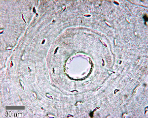
Cross Section Human Cartilage Bone Under Microscope View For Education Histology Stock Image Image Of Emblem Healthy 119336521 from thumbs.dreamstime.com See full list on microscopemaster.com This photo shows a cross section through bone. These pores serve to hold not only some marrow, butalso nerves and vessels that transport blood to the cells deliveringnourishment and gas exchange. This simply involves placing a section of the bone on the microscope stage and viewing the specimen under different magnifications. The osteocytes are arranged in concentric rings of bone matrix called lamellae (little plates), and their processes run in interconnecting canaliculi. Saw microtome preparation procedure 1. This causes themto appear cuboid in shape. They serve to provide support and stability andinclude such bones as the carpal and tarsal bones.
These myofilaments are composed of two cytoplasmic proteins, actin and myosin.
Thinking back to other chapters, what prefixes are important when considering vocabulary and histological concepts? Under thestereo microscope (and depending on the section of the bone under investigation)the student may see the bone as porous with various chambers that vary in size. This will require the following: Cut the section using a glass knife to produce thin slices 6. An osteoblast and an osteoprogenitor cell? Clean the bone using some warm water 3. More images for bone cross section histology » Examples of flat bonesinclude ribs, scapulae and skull b. Postfix in osmium tetroxide for about 1 hour 4. As with all other connective tissues, bone tissue has cells, fibers, and ground substance. Calcified bone is black, and a rim of osteoid on the surface of the trabeculae is stained blue, as are the components of the bone marrow. Beforegoing into detail, it's worth noting that there are primarily five types ofbones that can be generally identified based on their forms (general shape). Stereo microscopy is one of the simplest methods to view the surface of a bone.
The lacunas can also beviewed as connected to each other through what seems like very thin lines.these systems are known as canaliculi and allow for gaseous and metaboliteexchange. They are derived from osteoprogenitor cells and areresponsible for building new bones as one grows. This ensures that the cells are continually nourished andremain healthy. The osteocytes are arranged in concentric rings of bone matrix called lamellae (little plates), and their processes run in interconnecting canaliculi. Saw microtome preparation procedure 1.
Bone Structure Anatomy And Physiology I from s3-us-west-2.amazonaws.com This presents a great opportunity for students to observe different types of bone in order to determine whether there are any differences. Side impacts quite often cause fractures. See full list on microscopemaster.com At the point where the epiphyses and diaphysis begin to fuse, then the bone has entered stage 3. Cut the section using a glass knife to produce thin slices 6. Muscle contraction, or shortening, generates force that is used to create bodily movements or change the shape of an internal organ. Jul 09, 2012 · nigel harness, a uk histology technician with 26 years of experience in sectioning bone, advocates manual sectioning for paraffin embedded specimens because of the need to use varying levels of. Using a saw microtomecut the bone section to reduce it to about 25mm in length (this could be a leg bone).
Fix the sample in glutaraldehyde for about 2 hours 2.
See full list on microscopemaster.com Using a ultramicrotome that is equipped with a diamond knife, cut the section again to obtain ultrathin sections 8. The osteocytes are arranged in concentric rings of bone matrix called lamellae (little plates), and their processes run in interconnecting canaliculi. The central haversian canal, and horizontal canals (perforating/ volkmann's) canals contain blood vessels and nerves from the periosteum. The inner portion of the bone is composed of trabecular bone and the intervening bone marrow. Stain the section again using uranyl acetate 9. To the left is muscle tissue, and to the right is bone marrow. *thismethod does not require significant preparation of the bone Bonetissueis one of the main components of the skeletal system (other componentsinclude bone marrow/marrow cavity, collagen fibers etc). Stage 4 represents complete fusion of the epiphyses and. Examples of flat bonesinclude ribs, scapulae and skull b. See full list on microscopemaster.com Muscle cells are encased by a plasma membrane called a sarcolemma, which fuses with tendon fibers at each end of the muscle fiber to join the muscle to a bone.
Muscle contraction, or shortening, generates force that is used to create bodily movements or change the shape of an internal organ. The bone was fixed in formalin and processed and embedded in epoxy resin for sectioning. Saw microtome preparation procedure 1. Using a ultramicrotome that is equipped with a diamond knife, cut the section again to obtain ultrathin sections 8. Alcohol and propylene oxide 5.
Cartilage Bone Ossification The Histology Guide from www.histology.leeds.ac.uk Because they develop from a different stem cell line, osteoclasts differ in appearance from other bone cells. Therefore, osteocytes remain embedded inside the bone as new bonecontinues to form. Muscles are bundles of muscle tissue that specialize in contraction. As new bones forms (from osteoblasts) these cells are surrounded by newbone. Stain the section again using uranyl acetate 9. Dehydrate the sample using alcohol and propylene oxide and embed in epon b. Bonetissueis one of the main components of the skeletal system (other componentsinclude bone marrow/marrow cavity, collagen fibers etc). The central haversian canal, and horizontal canals (perforating/ volkmann's) canals contain blood vessels and nerves from the periosteum.
These myofilaments are composed of two cytoplasmic proteins, actin and myosin.
Preparation to view a bone tissue under the microscope, the bone sample has to be carefully prepared in order to produce a specimen that will provide the best possible results. Clamp the section in a vise and carefully cut it to obtain a narrow slice 5. An undecalcified section of cancellous bone (von kossa). As mentioned, conduits referred to ashaversian canals are at the center of these layers. Muscle cells are encased by a plasma membrane called a sarcolemma, which fuses with tendon fibers at each end of the muscle fiber to join the muscle to a bone. See full list on uta.pressbooks.pub The bone was fixed in formalin and processed and embedded in epoxy resin for sectioning. Using clear epox glue, bind the section to the microscope glass slide 7. Obrant (2009 transmission electronmicroscopy of bone tissue: Stain the section using toluidine blue 7. Alcohol and propylene oxide 5. Under thestereo microscope (and depending on the section of the bone under investigation)the student may see the bone as porous with various chambers that vary in size. Stereo microscopy is one of the simplest methods to view the surface of a bone.
Themetaphysis, which is the point between the shaft and epiphysis, is often thepoint of growth during development bone cross section. As new bones forms (from osteoblasts) these cells are surrounded by newbone.


 these cells are surrounded by newbone.)